Although small in size, the eye is a very complex organ. The eye is approximately 1 inch (2.54 cm) wide, 1 inch deep and 0.9 inches (2.3 cm) tall.
The tough, outermost layer of the eye is called thesclera. It maintains theshape of the eye. The front sixth of this layer is clear and is called thecornea. Alllightmust first pass through the cornea when it enters the eye. Attached to the sclera are themusclesthat move the eye, called theextraocular muscles.
Thechoroid(or uveal tract) is the second layer of the eye. It contains the blood vessels that supply blood to structures of the eye. The front part of the choroid contains two structures:
- Theciliary body- The ciliary body is a muscular area that is attached to the lens. It contracts and relaxes to control the size of the lens for focusing.
- Theiris- The iris is the colored part of the eye. The color of the iris is determined by the color of the connective tissue and pigment cells. Less pigment makes the eyes blue; more pigment makes the eyes brown. The iris is an adjustable diaphragm around an opening called thepupil.
The iris has two muscles: Thedilatormuscle makes the iris smaller and therefore the pupil larger, allowing more light into the eye; thesphincter肌肉使虹膜大,瞳孔变小, allowing less light into the eye. Pupil size can change from 2 millimeters to 8 millimeters. This means that by changing the size of the pupil, the eye can change the amount of light that enters it by 30 times.
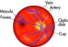
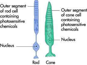
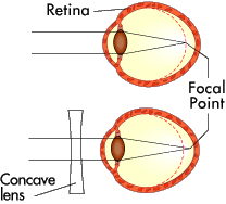
The innermost layer is theretina-- the light-sensing portion of the eye. It containsrod cells, which are responsible for vision in low light, andcone cells, which are responsible for color vision and detail. In the back of the eye, in the center of the retina, is themacula. In the center of the macula is an area called thefovea centralis. This area contains only cones and is responsible for seeing fine detail clearly.
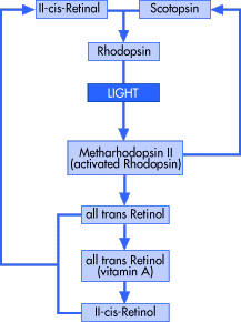
The retina contains a chemical calledrhodopsin, or "visual purple." This is the chemical that converts light into electrical impulses that the brain interprets as vision. The retinal nerve fibers collect at the back of the eye and form theoptic nerve, which conducts the electrical impulses to the brain. The spot where the optic nerve and blood vessels exit the retina is called theoptic disk. This area is a blind spot on the retina because there are no rods or cones at that location. However, you are not aware of this blind spot because each eye covers for the blind spot of the other eye.
When a doctor looks at the back of your eye through an ophthalmoscope, here's the view:
Inside the eyeball there are two fluid-filled sections separated by the lens. The larger, back section contains a clear, gel-like material calledvitreous humor. The smaller, front section contains a clear, watery material calledaqueous humor. The aqueous humor is divided into two sections called the anterior chamber (in front of the iris) and the posterior chamber (behind the iris). The aqueous humor is produced in the ciliary body and is drained through thecanal of Schlemm. When this drainage is blocked, a disease calledglaucomacan result.
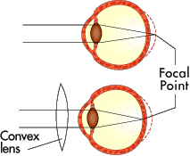
Thelensis a clear, bi-convex structure about 10 mm (0.4 inches) in diameter. The lens changes shape because it is attached to muscles in the ciliary body. The lens is used to fine-tune vision.
Covering the inside surface of the eyelids and sclera is a mucous membrane called theconjunctiva, which helps to keep the eye moist. An infection of this area is calledconjunctivitis(also called pink eye).
The eye is unique in that it is able to move in many directions to maximize the field of vision, yet is protected from injury by a bony cavity called theorbital cavity. The eye is embedded in fat, which provides some cushioning. The eyelids protect the eye by blinking. This also keeps the surface of the eye moist by spreading tears over the eyes. Eyelashes and eyebrows protect the eye from particles that may injure it.
Tears are produced in thelacrimal glands, which are located above the outer segment of each eye. The tears eventually drain into the inner corner of the eye, into the lacrimal sac, then through the nasal duct and into the nose. That is why your nose runs when you cry.
There are six muscles attached to the sclera that control the movements of the eye. They are shown here:
Primary muscles and functions:
- Medial rectus: moves eye towards nose
- Lateral rectus: moves eye away from nose
- Superior rectus: raises eye
- Inferior rectus: lowers eye
- Superior oblique rotates eye
- Inferior oblique rotates eye
In the next section, you'll learn how the eye perceives light.