How Brain Mapping Works

Neurons in the human brain as they transfer information among each other. Want a map of this? It could take a while. See morebrain pictures.
©iStockphoto.com/Kiyoshi Takahase Segundo

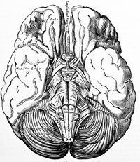
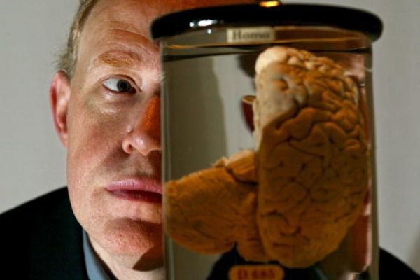
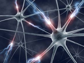
Thehuman brainis a knot of 100 billion neurons and supportcells. We can store a lifetime ofmemoriesthere. We can use it to write sonnets and buildairplanes. Sure, anelephant's brainis larger, weighs more, and has more neurons, but elephants also lack our abilities. Intrigued? Scientists sure are. That's one reason why they are mapping the human brain, a substantial project that could take decades to complete.

Brain mapping attempts to relate the brain's structure to its function, or finding what parts give us certain abilities. For example, what aspect of our brain allows us to be creative or logical? This is calledlocalization of function.

Advertisement

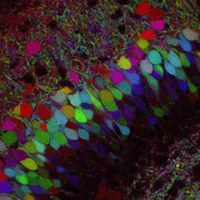
In mapping brain functions, scientists use imaging to watch the brain working on various tasks. Charles Wilson, a neurobiologist at the University of Texas at San Antonio, explains localization of function this way:

There's part of the brain that has to do primarily with vision and other parts that have to do primarily with sound. Now, can we look in the vision section and say, Is there a special part of the brain that detects red objects and another that detects green objects? Or does the same area detect objects of both colors?

Brain mapping also looks from the outside in. It examines how our environment changes our brain's structure by studying, for instance, how the brain changes physically through the learning andaging processes. Brain mapping also examines what goes wrong physically in the brain during mental illnesses and other brain diseases.

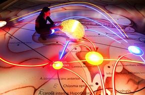
最后,大脑映射aims to give us a thorough picture of our brain's structure.Google Earthshows us satellite images ofour planetand zooms in to continents, countries, states, cities, highways, streets and buildings. A complete structural map of our brain might be similar. It could show us our whole brain; all the regions, functional lobes, specialized centers, thick neuron "bundles" connecting brain parts, neuron circuits, single neurons, junctions between neurons and finally, neuron parts. Scientists are still developing the parts that might form this massive map.

Brain mapping is a collection of many different tools. Researchers must collect images of the brain, turn those images into data, and then use that data to analyze what happens in the brain as it develops.

Read on to learn how researchers map the brain.

Advertisement

Brain Mapping Technology and Methods

We've come a long way from this.
Mark Strozier/©istockphoto.com

Scientists use many methods to study the brain's structure and function. They take pictures of healthy brains and compare them to diseased brains. In addition, they examine brains taken from humans,primatesand small mammals and try to understand how invertebrates' smaller nervous systems work. On a microscopic level, they also examine neurons.

Here are some tools used in brain mapping. These techniques take images of the brain:

Advertisement

  • Computer axial tomography (CAT) scanX-rays the brain from many angles and show structural abnormalities.
  • Structuralmagnetic resonance imagingtakes advantage of water in the brain to create images with better resolution than a CAT scan.
  • 扩散tensor-MRI (DTI)images "tracts" of neurons that connect brain regions by following water movement in the brain.

These techniques examine brain activity:

  • Electroencephalography (EEG)indicates electrically active locations in the brain using detectors implanted in the brain or worn on a cap.
  • Positron emission tomography(PET)takes images of radioactive markers in the brain.
  • Functional MRI (fMRI)shows images of brain activity while subjects work on various tasks.
  • Pharmacological functional MRI (phMRI)shows brain activity as drugs are administered.
  • Transcranial magnetic stimulation (TMS)noninvasively stimulates parts of the brain to trigger certain behaviors.

New methods allow researchers to see all the connections between neurons in an intact brain. This branch of study is calledconnectomics.The "wiring diagram" of a brain is called aconnectome[source:Lichtman]. "Until recently, we've had no hope of getting these wiring diagrams," says Jeff Lichtman, a Harvard biologist who led the group that developed some of the new techniques. "We could see individual cells, but never all of them at once."

Brainbow labeled the neurons in this transgenic mouse brain with about 90 different color combinations.
Stringer/Getty Images/AFP

One such technique, known asBrainbow, labels every neuron in a live animal's brain a different color. By generating images of the animal's brain, scientists can see where and how neurons connect to each other. As the animal grows and ages, they can also watch how the neurons change connections.

Another technique uses theATLUM,orautomatic tape-collecting lathe ultramicrotome. This machine reads the wiring diagram of a brain. "We do something akin to paring an apple," explains Lichtman. "We essentially shave off a spiral cut as we rotate the brain on a lathe and put this ribbon of tissue onto a tape. We'll eventually get a hugely long tape, which is essentially the whole brain. Using anelectron microscope, we will image that to see the structure of the wiring."

So far, Brainbow and the ATLUM are being used only to study animals with relatively small brains, like mice.

So, what's the point? What, if anything, can mapping accomplish? Learn what we can learn from mapping the human brain on the next page.

Advertisement

Uses of Brain Mapping

Why would scientists take on the arduous task of brain mapping? The answer is simple, says Lichtman: to understand our brains more intimately. We have never seen a diagram of how all of the neurons in the brain connect. As Jeff Lichtman puts it, "A lot of our thinking about the brain is based on incomplete knowledge of what is actually there. So we would like to see what is actually there."

The brain's wiring diagram may help us better understand how we learn and adapt, says Lichtman. "We start out being less well adapted to our environment than any other animal. By the time we're adults, we can use tools that our genetic heritage couldn't possibly have taught our nervous system to use -- like iPods. No other animal can do that. During our development, we must wire ourselves to [be able to] use these machines."

Advertisement

Brain mapping is also of practical use to doctors. Neurosurgeons use brain mapping to plan safer surgeries. One treatment for epilepsy, for example, removes the affected part of the brain. Using functional MRI and EEG, surgeons can locate the seizure center in a patient's brain -- as well as areas that are active during speaking and moving -- down to the millimeter. These images tell doctors what to leave and what to cut out.

Brain imaging is not only used in treatment. It is used to diagnose neurodegenerative diseases like Parkinson's and Alzheimer's [source: Wilson]. Using tagging techniques like PET, doctors look for drops in certain brain chemicals, or they may use MRI to examine shrinkages in areas show tissue loss. Over time, doctors can map what the brain looks like as diseases progress or as treatments work [source:Institute for Neurodegenerative Disorders].

Developmental disorders likeautismmay have a structural basis in the brain. Lichtman points out that autism is thought to involve a series of wrong connections between neurons. By applying Brainbow to a mouse with autism, researchers might see the wiring diagram evolve to find out how, when and if the wiring goes wrong.

Scientists have also sought to illustrate the effects of various mental illnesses in the brain, with some success. Brain imaging on these patients revealed structural abnormalities. For example, structural MRI has shown thatschizophrenicpatients lose matter in thetemporalandprefrontal cortexover time [Source:Rapoport]. These findings have yet to lead to treatments.

Panic disorder,bipolar disorder,depression, anxiety, eating disorders and more are being examined using different brain imaging techniques, but how do we interpret scientists' findings? More importantly, where can we see them? Find out on the next page.

Advertisement

The Brain Atlas: How We Use Brain Maps

Microsoft co-founder Paul G. Allen touts the completion of the Allen Brain Atlas on Capitol Hill in September 2006. The project studies the expression of genes in mouse brains.
Mark Wilson/Getty Images News

Neuroinformatics places all the data we have on the brain on theInternetin usable form. The data include images, models of neuron behavior and maps of thegenesthat are "turned on" in different brain regions. By making the data sharable and searchable, brain researchers can piggyback off of one another's studies and discover more.

Engineers are writing software to help brain researchers share and compare data. Software now analyzes, for instance, whether MRIs of Alzheimer's patients with different brain sizes and shapes have similar brain features. Are men with a certain brain architecture predisposed to bipolar disorder? This question, and many others, may one day be answered by computer programs that re-analyze images of past patients rather than by studying new ones.

Advertisement

Here are examples of brain atlases that researchers can mine for answers:

  • Allen Mouse Brain Atlas: The mouse brain expresses 21,000 genes. Researchers sliced up the brain and stained where each gene is expressed. Visitors can look at photos or3-D modelsof the brain for their gene of interest [source: Allen Institute for Brain Science].
  • Allen Human Cortex Atlas: Our cerebral cortex allows us to think, reason and remember. Researchers have sliced this up as well. They have also stained where 1,000 genes -- and counting -- are expressed. You can evensurfthe pictures [Source:Allen Institute for Brain Science].
  • Whole Brain Atlas: Stores images of the human brain as it ages and fights diseases [source:Becker].

Images aren't the only source of information. Here are examples of databases brain researchers use:

  • NeuronDBprovides diagrams of specific neurons in the human brain and tells you what inputs cause them to fire [source:Marenco].
  • ModelDBstores mathematical models of how neurons and neuron networks send electrical signals. "The collection of millions of cells doing that becomes movement, sensation, cognition, emotion, and human experience," says Wilson. "To make a model of the brain's function, we start with that" [source:Shepherd].

Now that we can map the human brain, how far have we gotten? Are we done yet? Are we even close? Find out on the next page.

Advertisement

The Complete Brain Map

A woman examines an exhibit in Dresden, Germany, showing the neurobiological processes of the human brain. Could we eventually map emotions like happiness?
Norbert Millauer/DDP

After imaging the brains of populations large enough to generate statistics, researchers have made sophisticated brain maps. There are maps to illustrate where we lose brain volume as we age, as AIDS progresses and as we use methamphetamines.

What would a complete map of the human brain look like? That depends on your interests. If you thirst to know the brain's structure, you might want to see that hypothetical Google Earth version that can begin with a picture of our cortex and zoom in to neuron number 888,898,432,857.

Advertisement

这个完整的,谷歌地球的地图是停滞不前at many points. One such point is the imaging of all of the human brain's neurons and their connections. Even getting this data in the mouse is painstaking, says Harvard biologist Jeff Lichtman. At the rate theATLUMand an electron microscope are now working, getting a map of all the interconnected neurons in the mouse brain would take 200,000 weeks, Lichtman estimates. The data would be "bigger than all the data on the Internet -- bigger than all the data in all the libraries in the world," he says. "At the moment, the kind of storage that's possible on computers is not quite up to task." The only "brain" for which we have a complete map of interconnecting neurons belongs toC. elegans, "a worm that's a millimeter long and has 300nervecells," says Lichtman.

Again, your definition of a complete brain map depends on your interests. If you're a neuropsychiatrist, for example, a complete map of the brain might be a time-lapse image showing how bipolar disorder unfolds in the brain from birth to the first symptom and what lithium does to stop the process.

That might not be enough for you. You might want to know the function of the brain's every last inch. Unfortunately, that's impossible. We can't capture functions that happen too quickly or too slowly, says neurobiologist Charles Wilson. Other processes take a lifetime. No imaging study has followed someone frombirthtodeath. "No method that we know of handles every time of interest. No method we know of handles more than a tiny piece of it," says Wilson. At this point, Lichtman says there is no current effort underway to integrate all of these maps into one.

But there's no fundamental reason why we can't eventually have any - or all -- of these maps, says Wilson. "The problems are all practical technology problems that can be overcome. As with any map, if you start with a crude map, it's better than no map. And you don't need a new map. You just add information to the old map to make it more refined. We aren't going to wake up one day and have this. We are going to add a little today, a little tomorrow, and at some point, we are going to say, 'Wow, this is starting to look pretty good.'"

For more information on the process of brain mapping, take a look at the next page.

Advertisement

Lots More Information

Related Articles

More Great Links

  • Allen Institute for Brain Science. "Human Cortex: Cortical Gene Expression." 7/15/2008. (7/16/2008) http://www.brain-map.org/content_description/content01
  • Allen Institute for Brain Science. "Allen Brain Atlas: Mouse Brain. 7/15/2008. (7/16/2008) http://mouse.brain-map.org/about/index.html
  • "Brain Scanning." Encyclopaedia Britannica. 2008. Encyclopaedia Britannica Online. (7/19/2008) http://www.search.eb.com/eb/article-9016179.
  • Bailer, Ursula. "Altered brain serotonin 5-HT1A receptor binding after recovery from anorexia nervosa measured by positron emission tomography and [carbonyl11C]WAY-100635." Archives of General Psychiatry. Vol. 62, no. 9. September 2005.
  • Becker, Alex and Keith Johnson. "The Whole Brain Atlas." (7/10/2008) http://www.med.harvard.edu/AANLIB/home.html
  • 布里尔,大卫。“谅解备忘录se Brain Map is Complete." Nature. Vol. 443, no. 7110. 9/28/2006.
  • Gura, Trisha. "Etiology of Eating Disorders Explored as Patience for a Cure Thins." Nature Medicine. Vol. 14, no. 5. May 2008.
  • Hurley, Dan. "The Science of Sarcasm (Not That You Care)." The New York Times. June 3, 2008. (7/9/2008) http://www.nytimes.com/2008/06/03/health/research/03sarc.html?_r=1&scp=1&sq=sarcasm%20mapped%20in%20the%20brain&st=cse&oref=slogin.
  • Institute for Neurodegenerative Disorders. "Functional Brain Imaging." (7/9/2008). http://www.indd.org/research/functional.html
  • Lichtman, Jeff. "A technicolor approach to the connectome." Nature Reviews Neuroscience. Vol. 9. 6/2008.
  • Lichtman, Jeff. Personal interview. Conducted 7/9/2008.
  • Matthews, Paul. "Applications of fMRI in translational medicine and clinical practice." Nature Reviews Neuroscience. Vol. 7, no. 9. 9/2006.
  • Marenco, Luis et al. "Neuron DB." (7/10/2008) http://senselab.med.yale.edu/neurondb/default.asp.
  • Murray, Graham. "Substantia nigra/ventral tegmental rewardprediction error disruption in psychosis." Molecular Psychiatry. Vol. 13, no. 3. 3/2008.
  • National Institute of Mental Health Child Psychiatry Branch. "Child Psychiatry Branch." 6/27/2008. (7/9/2008) http://intramural.nimh.nih.gov/chp/index.html.
  • Rapoport, Judith. "Brain neuroplasticity in healthy, hyperactive and psychotic children: Insights from Neuroimaging." Neuropsychopharmacology Reviews. Vol. 33, no. 1. 9/12/2006.
  • Shepherd, Gordon. "The Human Brain Project: neuroinformatics tools for integrating, searching and modeling multidisciplinary neuroscience data." Trends in Neurosciences. Vol. 21, no. 11. 11/1998.
  • Shepherd, Gordon. "Model DB." (7/10/2008) http://senselab.med.yale.edu/modeldb/default.asp.
  • Snyder, Solomon. "A Complex in Psychosis." Nature. Vol. 452, no. 7183. 3/6/2008.
  • Toga, Arthur. "Towards multimodal atlases of the human brain." Nature Reviews Neuroscience. Vol. 7, no. 12. 12/2006.
  • Wilson, Charles. Personal interview. Conducted 7/9/2008.
  • Zimmer, Carl. "What if There is Something Going on in There." The New York Times Magazine. September 28, 2003. (7/9/2008) http://query.nytimes.com/gst/fullpage.html?res=9503E0D71E3AF93BA1575AC0A9659C8B63.

Advertisement

Loading...