How Bones Work

By:Tom Scheve

A diagram showing back and side views of the human skeleton, circa 1900
赫尔顿存档/Getty Images
A diagram showing back and side views of the human skeleton, circa 1900

The human body is an incredible machine. It runs so well most of the time that we don't have to pay much attention to any of the life-sustaining systems that are in motion around the clock, humming along without our mindful involvement.

Right now, your body is performing vital and complicated tasks nearly too numerous to comprehend. Fortunately, our bodies don't demand our comprehension in order to pump theheart, oxygenate blood, regulate hormone production, interpret sensory data and carry out every other process that keeps our biological boats afloat.

In this article, we'll discuss one of the systems that makes life possible: the skeletal system.

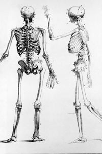
Bones prevent you from puddling on the floor in the form of a jellyfish, but what else do they do? Bones rebuild themselves, they producebloodcells, they protect ourbrainsand our organs, they provide a giant system of levers that allow us to move ourselves around, and bones also help maintain a steady amount of calcium in our bodies.

Bones and the Body

And, even if you never make your mark on the world (or in the history books), your bones will stick around long after you have otherwise vanished to declare to the world: "These skeletal remains once supported skin and tissue and organs! This person once existed!" And as the construction crew that unearthed your bones reels back in horror, every life choice you ever made will seem -- if but for a moment before you are shoveled into a Dumpster -- very much worthwhile.

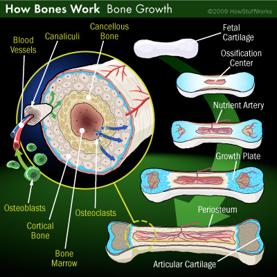
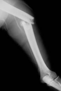
Before we leave behind our skeletal remains to freak out future generations, we should first learn some basics about bones: What are bones made of? What happens when they break? And just how many of them do you have, anyway?

Advertisement

Bone Basics

骨市场
没有什么可以阻止你合法purchasing human bones and skulls from a vendor in the United States. Specimens often come from China, where unclaimed remains are sold to bone distributors. Many skulls on the market today are from India, which was once a primary provider of skulls to the world market but banned the practice of exporting human remains in 1987. (Skulls don't come cheap: an average adult skull runs around $500.)

There are 206 bones in the adult body. Bone is a honeycomblike grid ofcalciumsalts located around a network of protein fibers. These protein fibers are calledcollagen.

When you patch a hole in a piece of drywall, you usually cover it with tape that has a gummy fibrous grid, and then cover that with wall compound mortar. Bone is made in much the same way. Collagen fibers are gummed together by a kind of shock-absorbing glue [source:加州大学ifornia-Santa Barbara]。Then, all of this is covered and surrounded by calcium phosphate, which hardens everything into place. Not only do bones make use calcium for strength, they also keep some stored in reserve. When other parts of the body need a calcium boost, the bones release the needed amount into the bloodstream.

There are two different types of bone tissue:cortical bone(the outer layer) andcancellous bone(the inner layer). Cortical bone, also known ascompact bone, provides external protection for the inner layer against external force. It makes up 80 percent of bone mass and is dense, strong and rigid [source:Hollister]。

Cortical bone is covered by a fibrous membrane called theperiosteum. Think of the periosteum as a utility vest that fits over the bone -- it has brackets and places formusclesand tendons to attach. The periosteum contains capillaries that are responsible for keeping the bone nourished withblood.

In the case of long bones such as the femur (the upper leg bone), the periosteum covers the central portion of the bone but -- like a sleeveless vest -- stops short of the cartilage tissue that resides on both ends of the bone (we'll discuss this cartilage in a later section).

Cancellous bone, also known astrabecularorspongy bone, is the inner layer of bone and is much less dense than cortical bone. It's formed by trabeculae, which are needlelike structures that create a meshwork. However, instead of a network of bone structure with periodic gaps, cancellous bone is more like a network of connecting spaces with periodic structure. The latticework of tiny chambers is filled either with bone marrow or connective tissue. Within these marrow-filled spaces is where new blood cells are produced.

Though cancellous bone only makes up about 20 percent of the body's bone mass, it plays important roles in body function. It provides structural stability and acts as a kind of shock absorber inside the bone, but without adding too much to the overall weight of the body.

In the next section, we'll learn more about bone marrow.

Advertisement

Bone Marrow

Inside the cavities of cancellous bone is soft, fatty tissue comprised of an irregular network ofbloodvessels and cell types. This is calledbone marrow. There are two types of marrow: red and yellow.

Red marrowcontains stem cells, unspecialized cells that can grow into different types of specialized cells. They're responsible for replenishing and replacing cells in the body that have been damaged or lost. (For the whole story on stem cells, give干细胞是如何工作的a read.) There are two types of stem cells found in red marrow:

  • Hematopoietic stem cells(HSCs).This type of stem cell is responsible for creating billions of new blood cells daily, at a rate of about 8 million every second [source:Houston Museum of Natural Science]。HSCs create every type of blood cell: red blood cells (which carry oxygen throughout the body), white blood cells (which fight infections and kill bacteria) and platelets (which help your blood clot). Marrow stem cells can even produce more marrow stem cells. HSCs can leave the marrow and enter the bloodstream, where the ratio of blood cells to stem cells is about 100,000-to-1 [source:National Institutes of Health]。

  • Stromal stem cells. This type of stem cell generates bone cells, cartilage, fat cells and connective tissue. Stromal stem cells are being studied for their possible use in repairing spinal cord damage and healing disorders of the lymphatic system.

Yellow marrowis mostly fat, and as weage, it can be found in places where red marrow once resided -- some of the bones in our arms, legs, fingers and toes, for instance. If the body needs more blood cells, yellow marrow can transform back into red marrow and produce them. Some bones have a lot more red marrow than others -- the pelvic bone, the spine's vertebrae and our ribs are all rich with it. The body also stores iron in bone marrow.

Bone marrow can become diseased. Myeloproliferative disorders (MPDs) cause the overproduction of immature cells from the marrow. Disorders such as aplastic anemia and myelodysplastic syndromes (MDS) hinder the marrow's ability to produce enough blood cells.

Several marrow diseases can be treated through stem celltransplants, which introduce healthy stem cells to the patient's body to replace the diseased cells. The traditional way to transplant these stem cells is to extract bone marrow from the donor's hip bone with a syringe and introduce the material into the recipient's body. You don't have to actually experience someone penetrating the process to imagine how unpleasant it is. Increasingly, doctors are harvesting the marrow stem cells from the bloodstream instead, resulting in better stem cell samples for the recipient and less pain and discomfort for the donor.

In the next section, we'll examine some of the bones that help prevent yourbrainand lungs from sliding down into your socks -- the axial bones.

Advertisement

Axial Bones

Bones can be broadly divided into two categories: axial and appendicular. In this section, we'll take a look at theaxialbones, so named because they form the axis of the body. Axial bones are associated with the central nervous system and protect delicate organs such as yourheartandbrain.


Axial bones include:

  • The cranium. Though that coconut on top of your neck feels like one big unit with a jaw attached, the cranium is actually comprised of 22 interlocking cranial and facial bones. These cranial plates and oddly shaped bones are held together by joints, though these joints (quite wisely) don't allow for movement (except for the mandible, or jawbone). Deep in your ear is the smallest bone in your body, the stirrup. It's about the size of a grain of rice.

  • human skeleton
    The spine
    . Your spine (also known as the vertebral column) is comprised of 33 specialized bones calledvertebrae. These vertebrae provide form for the rest of the body and protect the spinal cord. Starting from the head and moving downward, the first seven vertebrae arecervical vertebrae, which keep your beautiful cranium from rolling down the street every time you come to a sudden stop. They also allow you to nod "yes" or shake your head "no." Next are the 12thoracic vertebrae, forming the back of your rib cage. Below the thoracic vertebrae are thelumbar vertebrae, which bear much of the body's load. Most backmusclesare connected to these workhorses. Below these is thesacrum, which actually begins in childhood as five different vertebrae, but over time fuses into one unit. Below this is another single unit that begins life in several pieces, thecoccyx(tailbone).

  • The sternum. The sternum, or breastbone, is front and center in its role as organ-protector. It defends your heart,lungsand portions of your major arteries from external forces. Like the coccyx or sacrum, the sternum starts off as different sections that fuse over time into one unified piece. The sternum provides stability to the ribs that are attached to it.

  • The ribs. These flat bones form a protective shield around your internal organs. There are 24 ribs, 12 on each side of your body. They come in three different types. From the top, the first seven sets of ribs aretrue ribs. They connect in the back to the spine and connect in the front to the sternum. Next are thefalse ribs. These three pairs connect in the back to the spine, but in the front they attach to the seventh true rib, which is the last rib that connects to the sternum. Last are thefloating ribs, and these two pairs of ribs are attached to the spine like all the others, but "float" in the front without being attached to the sternum or any other rib.

In the next section, we'll learn about the bones that serve more than they protect: appendicular bones.

Advertisement

Appendicular Bones

而轴向骨骼形成th的纵轴e body, theappendicular bonesare the bones that connect to this axis. Unlike axial bones, protection isn't the function of appendicular bones; they're made for action. Let's take a look:

  • Bones of the shoulder. The bones that make up your shoulder girdle serve to connect your arms to your sternum and rib cage for stability and support. You have two clavicles (collarbones) that attach on one end to the breast plate and, on the other end, support the shoulder blades, or scapulas. The shoulder blades provide points of contact and attachment for manymusclesand the bone of each upper arm.

  • Bones of the arm and hand. The entire arm appendage has three basic components: the upper arm, the lower arm and the hand. The upper arm is onelong bone, the humerus. The top fits neatly into the scapula, and the lower end is connected by the elbow joint to the two bones of the lower arm: the ulna (the bone on the same side as your little finger) and the radius (the bone on the side of your thumb). The radius plays a larger role in your overall mobility and function, while your ulna provides more stability. Both the ulna and the radius connect to the wrist bones in the hand. Each hand has an impressive 27 bones: eight carpal bones that make up the wrist, five metacarpal bones that extend the length of your palm, and 14 phalanges that form four fingers with three bones each along with a single two-boned thumb.

  • The pelvic girdle. When you sit down, all the weight of your upper body rests ultimately on your pelvic girdle. This tough pair of hip bones protects lower organs such as the bladder and, for women, protects the development of a fetus and facilitatesbirth. The dimensions of the pelvic girdle differ fairly significantly for men and women, as the opening in the center of the girdle must be large enough for a child to pass through.

  • Bones of the thigh, leg and foot. Connecting the pelvic girdle to the lower leg is a bone in the thigh area called the femur, the longest and strongest in the body. About 25 percent of your total height is gained from the femur bone [source:Houston Museum of Natural Science]。通过膝关节股骨连接(范围h is covered and protected by the patella, or kneecap) to the shin bone (tibia). Slightly smaller than the tibia is the other bone in the leg, the fibula. The fibula is responsible for muscular connections, while the tibia makes sure your foot and your knee don't get any farther apart from each other. Each foot has 26 bones: seven tarsal bones that make up the ankle, five metatarsal bones that make up the body of your foot (and play a significant role in supporting your body's weight), and 14 phalanges that form -- as is the case with your fingers -- four toes with three bones each with a big toe that has two bones.

Next, we'll look at some characteristics of different bone shapes.

Advertisement

The Long and Short (and Flat and Irregular) of It: Bone Shapes

The 206 bones in the human body can roughly be divided into four categories: long, short, flat and irregular.

  • Dr. Bess Mensendieck in 1995 teaching Body Mechanics
    Von Haven/BIPs/Getty Images
    Dr. Bess Mensendieck in 1995 teaching Body Mechanics
    Long bones. Not alllong bonesare actually long (some bones in the fingers and toes are long bones), but most are (such as the leg and arm bones). Long bones are identified by the shape and structure of the bone: a slightly curved shaft capped on both sides with hyaline cartilage, longer than it is thick. They're made mostly of compact bone, allowing them to support great amounts of weight and withstand pressure.

    The femur provides an excellent example of the strength of long bones. Its hollow cylindrical design allows it to provide the most possible strength out of the materials provided (the substance of the bone itself), without making it too heavy. The hollow internal portion of many long bones is where the marrow is located. Long bones grow from both ends, and have a cartilage plate (also known asepiphyseal plates) between the bone shaft and each bone end. These plates continue growing through adolescence.

  • Short bones. Short bones consist mainly of spongy bone with a protective covering of compact bone. Short bones are neither long nor thick, but rather cubelike. Your kneecaps (patellae), wrists (carpals) and some of the bones in yourfeetand ankles (tarsals) are short bones. Short bones aren't designed for a great deal of movement, but they're sturdy, compact and durable.

    The short bones in the wrist and ankle are also known assesamoid bones. Sesamoid bones (usually classified as short or irregular bones) are placed within tendons in parts of the body where a tendon must cross a joint. These bones hold the tendon slightly away from the joint to provide better range of motion when the tendon tightens. For instance, your kneecap connects two pieces of tendon that cross over the joint between your femur (upper leg bone) and the tibia of your lower leg.

  • Flat bones. These bones are thin and flat. Flat bones have a middle layer of spongy bone located between two protective layers of compact bone. These bones are generally protective in nature. Flat bones make up most of the 29 bones that fuse together to form the skull and protect thebrain, and also protect the major internal organs by forming the 24 ribs (12 on each side) of the rib cage, as well as the sternum. The shoulder blade (scapula) is also a flat bone. Flat bones contain marrow, but aren't large enough to have marrow cavities -- marrow is found instead in the spongy bone. The marrow in flat bones produces more redbloodcells than any other adult bone type.

  • Irregular bones. The bones that don't fit in the other three categories are irregular bones. Vertebrae in the spine and the jawbone (mandible) are irregular bones. This type of bone usually has a very specialized function and is made up of mostly spongy bone with a thin layer of compact bone around it.

In the next section, we'll learn that even the laziest among us is really a bone-making workaholic.

Advertisement

Bone Construction Zone: How Bones Grow

Human bone growth

Right now, the bones in your body are undergoing renovation. There are wrecking crews blasting into the bone quarry and carting off debris while an entirely different work crew hauls bags of concrete to the blast site and patches the newly made holes with stronger, newer, better material.

Before we talk about replacing bone with bone, we'd better learn how cartilage turns into bone. When you're floating around in the womb, your developing body is just beginning to take its shape, and it's creating cartilage to do so. Cartilage is a tissue that isn't as hard as bone, but much more flexible and, in some ways, more functional. Cartilage is pretty good stuff to use if you're going to mold a human -- good enough for the finer work, especially, such as your nose or your ear.

A large amount of thatfetuscartilage begins transforming into bone, a process calledossification. When ossification occurs, the cartilage (which doesn't have salts or minerals in it) begins to calcify; that is, layers of calcium and phosphate salts begin to accumulate on the cartilage cells. These cells, surrounded by minerals, die off. This leaves small pockets of separation in the soon-to-be-bone cartilage, and tiny blood vessels grow into these cavities. Specialized cells calledosteoblastsbegin traveling into the developing bone by way of thesebloodvessels. These cells produce a substance consisting of collagen fibers and they also aid in the collection ofcalcium, which is deposited along this fibrous substance. (One common analogy for this design is reinforced concrete, which is a grid of metal rods covered with concrete mix.)

After a while, the osteoblasts themselves become part of the mix, turning into lower-functioning osteocytes, sort of a retired version of osteoblasts that continue to putter along but don't stray too far from the blood vessels. This osteocyte network helps form the spongelike lattice of cancellous bone. Cancellous bone isn't soft, but it does look spongy. Its spaces help transfer the stress of external pressures throughout the bone, and these spaces also contain marrow. Little channels calledcanaliculirun all throughout the calcified portions of the bone, enabling nutrients, gases and waste to make their way through.

But we're not through with the bone-growing process yet.

Advertisement

Bone Growth, Continued

The Stretching of the Lambs
Baby lambs do about 90 percent of their growing at night, and the same may hold true for growing humans. This may be a primary reason why growing pains are experienced mostly at night by young children [source:英国广播公司]。

Before turning into osteocytes, osteoblasts produce cortical bone. One way to imagine this process is to picture a bricklayer trapping himself inside a man-sized brick chamber of his own construction. After forming the hard shell (cortical bone), the bricklayer himself fills the chamber. Air makes its way through the brick and decays the bricklayer. In bone, this part of the process is accomplished byosteoclasts, which make their way into the calcifying cartilage and take bone out of the middle of the shaft, leaving room for marrow to form. Osteoclasts do this by engulfing and digesting the bone matrix using acids and hydrolyticenzymes. So, our bricklayer (osteoblast) made the tomb (cortical bone), died inside the tomb (became an osteocyte), decayed over time (dissolved by osteoclasts) and left behind his remains that formed a network of mass and space inside the brick tomb.

最终,所有的软骨变成了骨头,except for the cartilage on the end of the bone (articular cartilage) and growth plates, which connect the bone shaft on each side to the bone ends. These cartilage layers help the bone expand, and finally calcify by adulthood.

So, right now in your body, there are osteoclasts hard at work absorbing old bone cells and osteoblasts helping to build new bone in its place. This cycle is calledremodeling. When you're young, your osteoblasts (the builders) are more numerous than the osteoclasts, resulting in bone gain. When you age, the osteoblasts can't keep up with the osteoclasts, which are still efficiently removing bone cells, and this leads to loss of bone mass (and a condition called osteoporosis, which we'll discuss shortly).

What good is making all this bone if you don't get to break some now and then? We'll further explore this line of questioning in the next section.

Advertisement

The Breaks: Bone Fractures

arm fracture
©iStockphoto.com/muratseyit
What happens when your sibling shoves you off the top bunk

Although bone is very strong material, it can break in a number of ways with enough force pushing, pulling or twisting it. Here are some of the more common breaks:

  • Stress fracture. Thistype of fractureis the result of sustained force on a bone, such as that created by running or jumping. Most stress fractures occur in the lower body, due to the accumulated weight that the bones in our legs and feet must support. It's possible to have a stress fracture without feeling any pain.

  • Open fracture. Unlikeclosed fracturesin which all portions of the broken bone remain within the skin, open fractures result in a piece of bone puncturing and piercing the skin.

  • Complete fracture. This is when the bone breaks neatly into two pieces.

  • Single fracture. This identifies a break in which the bone has only one damaged area.

  • Comminuted fracture. Also known as "More painkillers, please," comminuted fractures are bones that have been crushed or broken into more than two fragments.

  • Greenstick fracture. With this type of fracture, the bone has cracked on one side, but not all the way through. These breaks normally occur in children.

  • Pathologic fractures. These fractures may be caused by external forces, but the underlying cause is a bone that has been weakened by disease or infection, such as bone cancer.

  • Displaced fractures. The two broken ends of the bone don't line up and require repositioning before they're set in place.

  • Simple transverse. This type of fracture is an even, perpendicular break to the bone. (Imagine if someone chopped your femur bone in half by neatly striking it from the side at a right angle.)

  • Oblique fracture. An oblique fracture would be a diagonal fracture running lengthwise along the bone. (Think greenstick fracture, but all the way through the bone.)

  • Spiral fracture. Spiral fractures occur when the bone has been twisted past its maximum point of resistance.

Here are a few of the more common break locations:

  • Colles' fractureis just a fancy name for abroken wrist. It's also the most common type of fracture.

  • Hip fractures. More than half the fractures seen in older people arehip fractures[source:FDA]。Hip fractures are actually a break in the femur bone, right below the joint that connects the upper portion of the femur to the pelvis.

  • Compression fractures. This is a fracture that affects the spine. Bad falls are usually responsible for compression fractures, in which one or more vertebrae are essentially crushed.

Now that we know how bones break, let's see how they're mended.

Advertisement

Mending the Break: Healing Broken Bones

When bones are fractured, the body immediately initiates the first phase of healing, thereactive phase. Rupturedbloodvessels gather at the site of the break and form a clot. This clot containsfibroblasts, which are connective tissue cells that produce collagen proteins. When this clot forms, it lays the groundwork for what will be a full-scale restoration of the bone.

In a few days, the broken ends of the bone produce new blood vessels that grow into the clot that now bridges the separation in bone caused by the fracture. White blood cells arrive with these new blood vessels and begin carting away unneeded material from the site of the break. Now, the fibroblasts begin to multiply and secrete collagen fibers which form a matrix that replaces the blood clot.

In the修复阶段, specialized cells --osteoprogenitorcells -- located in the periosteal membrane that covers most of the bone begin transforming into different types of needed cells. Some of these cells --chondroblasts-- produce cartilage, while others -- osteoblasts -- produce uncalcified bone calledcallus. The new cartilage and callus bridge the separated pieces of bone, and the cartilage begins to ossify into trabecular bone.

In the third phase, theremodeling phase, osteoclasts begin removing the trabecular bone while osteoblasts replace it with compact bone. Once this phase is finished, the fractured bone has healed. (For more on the healing process, tryHow do broken bones heal?)

Sometimes whenbones break他们必须重新在骨折ds line up correctly for healing. All pressure must be taken off the bone, so that the ends that are attempting to fuse back together don't move out of position. Surgery might need to be performed to hold the bone fragments together with metal plates, rods or screws. Not only do these devices secure the bone's position for healing, they provide a starter bridge for the calcium deposits that will begin accumulating along the healing area.

Next, we'll learn about what happens when two bones meet in the night (or day).

Advertisement

Hooking Up: Joints

Each time you lean forward, pick up a cup of coffee, raise it to your lips and put it back down, your bones,joints,muscles和其他tissuesare all working in perfect synchronicity to make this effort possible.

There are 68 joints in the human body, and each joint is comprised of several elements. Among them are:

  • Bones. Well yeah, but more precisely, thearticular cartilageat the end of the bones. This cartilage prevents the bone ends from being damaged by contact with each other. Cartilage itself can be harmed by infection, injury, disease or simple wear and tear. This damage may lead to pain, inflammation and stiffness, a condition known asarthritis.

  • Skeletal muscle. Skeletal muscle attaches to bone and appears striped when viewed closely, earning the name "striated muscle." Unlike your cardiac muscle or the muscle in the walls of your stomach, skeletal muscle can be voluntarily moved, and lie at rest when not consciously activated. These muscles connect to bones through tendons.

  • Tendons. When skeletal muscle contracts or lengthens, it pulls on bone through an attached tendon, a tough, flexible tissue.

  • Ligaments. These tissues are pretty similar to tendons, except they connect bone to bone, ensuring that bones that meet together to form a joint will stay in place.

  • Synovial membrane.This layer of connective tissue exists around each joint, providing it with protection and producing synovia, a fluid that lubricates the joint and nourishes the cartilage.

  • Bursa. Similar to the synovial membrane, the bursa is a small sac that provides a lubricant to ease the movement of muscle against muscle or muscle against bone.

Not every joint moves. The skull, for example, consists of several bone plates that join together, but the fibrous tissue connecting these plates doesn't allow for movement.

Open, Sesamoid: Types of Joints

Freely moveablejointsfall into one of several different categories. The different types of joints are:

  • Pivot joints(known also asrotary joints).这些关节允许绕轴旋转。There is a pivot joint near the top of your spine that allows your head to move from side to side.

  • types of joints
    Hinge joints
    . This type of joint can open and close like a door. Your elbow is a hinge joint. Your biceps and tricepsmusclesare basically two people standing on opposite sides of a wall (the humerus, or upper-arm bone), each with one hand reaching over to its respective side of a door (the bones of the lower arm). The biceps "shuts" the door, by contracting and lessening the degree of the joint angle, and the triceps, when it pulls on its respective side of the door, "opens" the door, as the hinge then widens.

  • Gliding joints(known also asplane joints).This type of joint features two bone plates that glide against one another. The joints in your ankles and wrists are gliding joints. (Holding your forearm steady while your hand points upward and then waving side-to-side with your hand is an example of this joint's functioning.)

  • Ball-and-socket joints. This is the most maneuverable type of joint. Yourshoulderand your hip are both ball-and-socket joints. These joints feature a connection between one bone-end equipped with a protrusion that fits into the receptive space at the end of the other bone in the joint. These joints allow for forward motion, backward motion and circular rotation.
  • Saddle joints. These joints allow for two different types of movement. For instance, a saddle joint allows your thumb to move toward and away from your forefinger (as when you spread all five digits out, then bring them all together side-by-side) as well as cross over the palm of your hand toward your little finger.

  • Conyloid joints. These joints are similar to ball-and-socket joints, just without the socket (the "ball" simply rests against another bone end).

Next, we'll talk about what happens when bones go bad.

Bone Loss

woman drinking milk
©iStockphoto.com/evemilla
Give your thirsty bones the calcium they need -- and a fighting chance against osteoporosis.

Bones, like any other part of the body, are susceptible to disease, the most common beingosteoporosis.Osteoporosisis the diminishing of bone mass, leaving it structurally brittle and physically porous. One in six Americans has osteoporosis or early signs of the disease [source:National Institute of Arthritis and Musculoskeletal and Skin Diseases]。In especially serious cases, a bone can be broken by as little as a sneeze.

Women are greatly affected by this condition -- four out of five cases of osteoporosis occur in women, and half of all women over the age of 50 will have a fracture related to osteoporosis [source:National Osteoporosis Foundation]。But it also affects men and the young -- in fact, one quarter of all men over 50 will suffer an osteoporosis-related fracture. Though any bone can be affected by osteoporosis, the hip, spine and wrist are the most common. Fractures of the hip and spine are especially problematic, resulting in immobility, severe and lasting pain and even death.

Contributing factors of osteoporosis include:

  • Sedentary life. It's important to getexercise, since any weight-bearing activity will improve the strength of your bones. Additionally, exercise will prompt glands in the body to produce hormones -- such as growth hormone, testosterone or estrogen -- that help prevent the deterioration of your bones as you age. Other lifestyle choices, such assmokingand possibly highalcoholintake, adversely affect bone mass.

  • Malnutrition. A poor diet will result in not getting enough vitamin C,calcium, phosphorus, magnesium andvitamin D-- all important elements of good bone health and the sustained ability of your bones to produce new bone matrix. Talk to your doctor about your diet and vitamin intake.

  • Underdevelopment of bone mass before the age of 20. You most likely will have achieved the majority of your peak bone density by about age 20, though you can still gain bone mass until you're 30 or so. Living a healthy lifestyle and gaining bone mass early on will pay dividends down the road as you age.

  • Low estrogen. Women with higher estrogen levels tend to have higher bone density.

Other Bone Diseases

Osteoporosiscan lead to a condition calleddowager's hump. This occurs when the spine, due to diminished mass and strength, begins to compress, resulting in an outward curve of the upper vertebrae in the spine.

Another disease that can affect the bones isbone cancer. Bonecancermost often spreads to the bone from other parts of the body, but it can also start in the bone. When it begins in the bone, it's known asprimary bone cancer. Fortunately, primary bone cancer is pretty uncommon -- there are about 2,300 new cases discovered each year [source:National Cancer Institute]。When found, bone cancer can be treated by surgically removing thetumorfrom the bone or using chemotherapy, radiation therapy or cryosurgery (killing cancerous cells by freezing them with liquid nitrogen).

Osteonecrosisis a condition in which bones no longer receive thebloodthey need to survive, leading to bone death and degeneration. The cause of the disease isn't currently known. Most cases require surgical intervention, and doctors can graft healthy bone onto the diseased portions, attempt to restore blood flow or replace joints with mechanical joints.

Osteogenesis imperfectais an inherited disease that causes bones to be especially brittle. A faulty gene leaves the body unable to produce collagen normally. (For more, readHow Osteogenesis Imperfecta Works.)

Paget's disease of boneaffects about 1 million people in the United States and tends to show up more often in people with Northern European ancestry [source:National Institute of Arthritis and Musculoskeletal and Skin Diseases]。Paget's disease of bone affects seemingly random bones in the body, making them grow too large and structurally unstable. While the disease can affect any bone, it most often affects the pelvis, skull, spine and leg bones.

See the next section for lots more information about bones and the human body.

Lots More Information

Related HowStuffWorks Articles

More Great Links

Sources

  • American Association for Clinical Chemistry. "Myeloproliferative Disorders." June 27, 2005. http://www.labtestsonline.org/understanding/conditions/myelopro_disorders.html
  • Bridwell, Keith, MD. "Basic Bone Structure." Jan. 17, 2008. (Jan. 15, 2008) http://www.spineuniverse.com/displayarticle.php/article1224.html
  • 英国广播公司. "Bones 'grow most at night time.'" Jan. 31, 2005. http://news.bbc.co.uk/1/hi/health/4215003.stm
  • CIA. "The World Factbook: United States." Jan. 22, 2009. https://www.cia.gov/library/publications/the-world-factbook/print/us.html
  • Emuseum. "Human Anatomy." (Jan. 22, 2009) http://www.mnsu.edu/emuseum/biology/humananatomy/
  • Hendry, Joene. "Teen depression, smoking hard on the bones." Reuters Health. Dec. 12, 2008. http://www.nlm.nih.gov/medlineplus/news/fullstory_72722.html
  • 霍利斯特,斯科特·J。博士。“骨骼结构”。(1月。24, 2009) http://www.engin.umich.edu/class/bme456/bonestructure/bonestructure.htm
  • Hou, Chuanqiang; Wu, Xuejun; Jin, Xing. "Autologous Bone Marrow Stromal Cells Transplantation for the Treatment of Secondary Arm Lymphedema: A Prospective Controlled Study in Patients with Breast Cancer Related Lymphedema." Sept. 5, 2008. http://jjco.oxfordjournals.org/cgi/content/abstract/38/10/670
  • Houston Museum of Natural Science. "Body Facts." Jan. 23, 2009. http://www.hmns.org/exhibits/special_exhibits/bodyworlds/bodyworlds_body_facts.asp?r=1
  • Mayo Clinic. "Greenstick fractures." Oct. 21, 2008.
    http://www.mayoclinic.com/health/greenstick-fractures/DS00817
  • MedlinePlus. "Bone Diseases."
    http://www.nlm.nih.gov/medlineplus/bonediseases.html
  • Joseph, Thomas N., M.D. "Flat bones." Aug. 6, 2007 (Jan. 21, 2009) http://www.nlm.nih.gov/medlineplus/ency/article/002366.htm
  • MedlinePlus. "Long bones."
    http://www.nlm.nih.gov/medlineplus/ency/article/002249.htm
  • Merck. "Fractures." Feb. 2003. (Jan. 20, 2009) http://www.merck.com/mmhe/sec05/ch062/ch062a.html
  • National Cancer Institute. "Bone Cancer." (Jan . 22, 2009) http://www.cancer.gov/cancertopics/factsheet/sites-types/bone
  • National Cancer Institute. "Skeletal System." (Jan. 22, 2009) http://www.nlm.nih.gov/medlineplus/ency/article/002249.htm
  • National Institute of Arthritis and Musculoskeletal and Skin Diseases. "Paget's Disease of Bone." Jan. 2008. (Jan. 22, 2009)
  • http://www.niams.nih.gov/Health_Info/Bone/Pagets/default.asp
  • National Institutes of Health. "Stem Cell Information." Sept. 15, 2008. http://stemcells.nih.gov/index.asp
  • National Osteoporosis Foundation. Jan. 22, 2009.
    http://www.nof.org/
  • National Space Biomedical Research Institute. "Human Physiology in Space: Skeletal System." (Jan. 22, 2009)
    http://www.nsbri.org/HumanPhysSpace/focus6/ep_intro.html
  • Oklahoma Surgical Hospital. "Types of Joints." (Jan. 21, 2009) http://www.orthooklahoma.com/clinical/jointtypes.html
  • OSF HealthCare. "Bone fractures." (Jan. 21, 2009) http://stayinginshape.com/3osfcorp/libv/r04.shtml
  • Reeser, Jonathan C, M.D., Ph.D. "Stress Fracture." Feb. 21, 2007. http://emedicine.medscape.com/article/309106-overview
  • 加州大学ifornia - Santa Barbara. "Fundamental Discovery About The Fracture Of Human Bone: It's All In The 'Glue'." ScienceDaily 20 July 2005. 15 January 2009 http://www.sciencedaily.com /releases/2005/07/050718214954.htm
  • WebMD. "Colles' Fracture (Distal Radius Fracture)." Feb. 1, 2007. (Jan. 21, 2009) http://www.webmd.com/a-to-z-guides/colles-fracture

Games

Advertisement

Loading...